Life Science
Hard tissue
High resolution IR and Raman Imaging of hard tissue matter
Sub-micron IR analysis of tooth lateral cross-section
- Despite rough topography, dispersion-artefact free, spatially resolved submicron IR spectra and IR chemical images showing the chemical difference between the enamel vs. the dentin could be obtained in reflection mode off thick slices of teeth.
- Protein absorption are more prominent in the dentin while the enamel has stronger phosphate absorption from hydroxyapatite
IR Polarized O-PTIR to study collagen orientation in individual fibrils and tendon
A: Spectra obtained with O-PTIR from control tendon fibrils on CaF2 window. B: Single frequency image at right recorded at 1655 cm-1 in perpendicular orientation. markers denote locations at which spectra were acquired. Scale bar = 1µm
C and D: Optical photothermal IR (O-PTIR) spectra from intact tendon, from ~500 nm measurement spots. (B) Individual spectra obtained from the two orientations of a section mounted on a CaF2 window, relative to the linearly polarized QCL. Inserted visual image shows the 6 locations, all of which lie within the region imaged with FTIR FPA; scale bar = 70 μm.
Colored markers (+) correspond to spectral colors. (C) Comparison of spectra obtained from CaF2 (top) and glass (bottom) substrates in parallel and perpendicular orientations to linearly polarized QCL.
Published: Gorker Bakir et al., “Orientation Matters: Polarization Dependent IR Spectroscopy of Collagen from Intact Tendon Down to the Single Fibril Level”, Molecules 2020, 25, 4295
Widefield O-PTIR measurements on autofluorescent collagen fibril
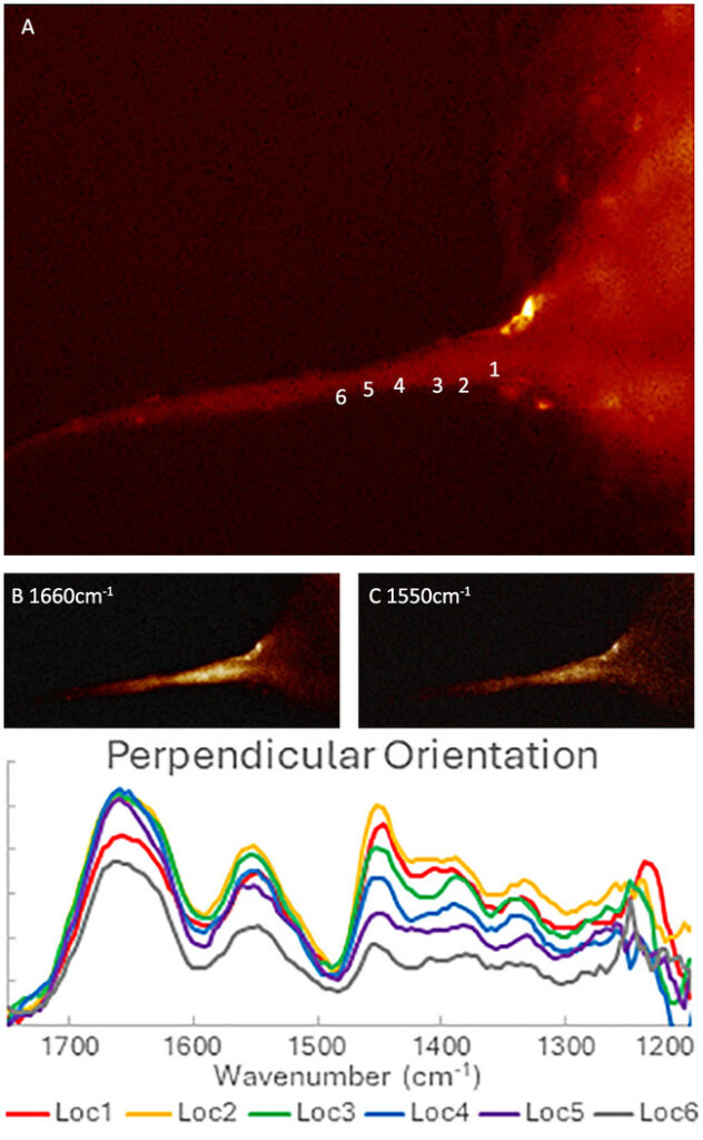
FL-PTIR measurements on an autofluorescent collagen fibril at different orientations. (A) Autofluorescent emission image of collagen fibril excited at 365 nm. (B-C) FL-PTIR absorption images at 1660 cm-1, 1550 cm-1, respectively. (D) FL-PTIR spectra extracted from an IR hyperspectral image stack from the indicated regions of interest for IR radiation oriented substantially perpendicular (E) to the fibril.
(Sample courtesy of Prof. K Gough, University of Manitoba).
Webinars
- Life Science
Alzheimer’s Disease Research with Sub-micron Simultaneous IR+Raman: Co-localization of Beta-Sheets and Carotenoids in Aggregates
- October 30, 2024
- Life Science
Life science applications using novel submicron simultaneous IR and Raman microscopy – A new paradigm in vibrational spectroscopy
- October 3, 2019
- Life Science
Amyloid aggregates in neurons – Life science applications using submicron simultaneous IR and Raman microscopy
- March 26, 2020
- Life Science
Collagen orientation, fiber to submicron fibril life science applications of O-PTIR, from tissues to single cells and bacteria
- January 22, 2021
- Life Science
Single and intra-cell bacterial IR spectroscopy
- March 10, 2021
- Life Science
Live single cell analysis with simultaneous submicron IR+Raman spectroscopy
- April 29, 2021
Need more information?
Discover how O-PTIR technology can elevate your research or help solve your toughest challenges. Our team are happy to assist and answer your questions.